
Articles

Delta Vista Optometry Blog

Delta Vista Optometry Blog is your trusted source for informative articles, expert insights, and valuable tips related to eye health and vision care. Explore a wealth of knowledge on topics ranging from eye conditions, vision correction options, to maintaining optimal eye wellness. Stay informed and prioritize your eye health with our engaging and educational blog content.

Blephex

Blephex

Our eyes are extremely delicate, yet they can be subjected to harsh conditions and other environmental factors that affect their health. One of the problems that can affect our eyes is an accumulation of dirt, debris and bacteria on the eyelids. This can cause a range of issues, including stopping tear film from reaching the eyes and being properly dispersed over their surface – which is necessary to keep them healthy and comfortable. Fortunately, a new solution called Blephex® can help.
 

What is Blephex®?

Blephex® is a handheld electro-mechanical device that is applied to the margins of the eyelids with the purpose of cleaning them and improving the effectiveness with which tear film flows onto the surface of the eyes.

Blephex® has a disposable, surgical-grade sponge tip which rapidly oscillates to create a cleaning action. Before the sponge tip is placed onto the eyes, it is soaked in a gentle exfoliating solution. This solution provides soft abrasion to help remove dead skin cells and debris that could be irritating the eyes and interrupting tear film progression. The Blephex® device is manually applied to the eyes and moved gently across the eyelids, with the entire, painless process taking approximately 6 to 8 minutes per eye. A different sponge is used on each eye, ensuring that no bacteria is passed between them. After the procedure, patients are given instructions on how to maintain the cleanliness of their eyelids with daily/nightly eyelid hygiene at home.

Most patients experience a significant improvement in tear film production and dispersal, and a reduction in unpleasant symptoms that they may have been experiencing within 48 hours of their treatment. While a single treatment is normally enough to produce excellent results, many patients are advised to have Blephex® every 4-6 months.

Read more
Low Level Light Treatment

Low Level Light Treatment

Dry eye is a common condition that occurs when the eyes do not produce enough tears or when the tears evaporate too quickly. There are several factors that can contribute to dry eye, including environmental factors, hormonal changes, and certain medications. Additionally, conditions such as meibomian gland dysfunction and blepharitis can also lead to dry eye.
 

The Role of Meibomian Gland Dysfunction and Blepharitis in Dry Eye

Meibomian gland dysfunction occurs when the meibomian glands, which are responsible for producing the oily layer of tears, become blocked or do not function properly. This can result in the tears evaporating too quickly and not providing enough lubrication for the eyes. Blepharitis is an inflammation of the eyelids that can cause redness, itching, and a gritty sensation in the eyes. Both meibomian gland dysfunction and blepharitis can contribute to dry eye by disrupting the natural tear film.
 

Recognizing the Symptoms of Dry Eye

Dry eye can cause a variety of symptoms, which can vary in severity from person to person. Some common symptoms of dry eye include:

  • Dryness: The most common symptom of dry eye is a feeling of dryness or grittiness in the eyes. This can make it uncomfortable to wear contact lenses or spend long periods of time looking at a screen.

  • Redness: Dry eye can cause the blood vessels in the eyes to become more prominent, leading to redness and irritation.

  • Itching: Some people with dry eye may experience itching or a burning sensation in their eyes.

  • Excessive tearing: Dry eye can also cause excessive tearing. This is the body's response to the irritation caused by the lack of lubrication in the eyes.
     

If you are experiencing any of these symptoms, it is important to consult with an optometrist for a proper diagnosis and treatment plan.

Read more
LACRIFILL® (Punctal Plugs)

LACRIFILL® (Punctal Plugs)

For many individuals, dry eye disease is an ongoing struggle marked by irritation, burning, watery eyes, and inconsistent vision. While over-the-counter drops may offer short-term relief, they often fail to provide lasting hydration. In many cases, the issue isn’t a lack of tear production - it’s that tears drain away too quickly.

One innovative option designed to provide longer-term relief is LACRIFILL®, a modern approach to punctal occlusion that helps preserve your natural tears and restore balance to the ocular surface. 
 

What Is LACRIFILL® Canalicular Gel? 

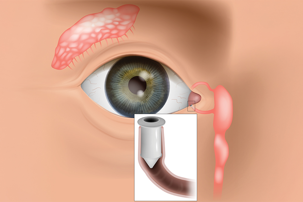
LACRIFILL® is a soft, clear gel that is placed inside the eye’s natural tear drainage channels, known as the canaliculi. These tiny channels are responsible for carrying tears away from the surface of the eye and into the nasal cavity. 

For individuals with dry eye disease, tears often drain away too quickly. When this happens, the eye does not stay properly lubricated, leading to dryness and irritation. LACRIFILL® works by gently filling part of the drainage pathway, slowing tear outflow so that your natural tears remain on the eye longer.

Unlike traditional artificial tears that supplement moisture from the outside, LACRIFILL® helps you retain the tears your body already produces. This treatment is FDA-cleared and has been clinically studied for safety and effectiveness in managing dry eye symptoms. 


How Does LACRIFILL® Work?

Every time you blink, a thin layer of tears spreads across the surface of your eye. This tear film keeps your eyes smooth, hydrated, and protected from debris and infection. After serving their purpose, tears drain through small openings in the inner corners of the eyelids (called puncta), passing into the canaliculi and eventually into the nose. In dry eye patients, this drainage process may occur too rapidly, leaving the surface of the eye exposed and under-lubricated. 

LACRIFILL® forms a soft gel barrier within the drainage system, reducing tear loss and improving moisture retention. By keeping tears on the eye longer, it supports a healthier tear film and enhances overall comfort. 

The gel gradually biodegrades over time and is naturally reabsorbed by the body. Relief typically lasts several months, and the treatment can be repeated when necessary. 
 

Benefits of LACRIFILL®

LACRIFILL® offers several advantages for patients struggling with persistent dry eye symptoms: 

  • FDA-cleared and clinically evaluated for safety and performance

  • Quick, non-surgical procedure performed in-office

  • No downtime or recovery period required

  • Preserves your body’s natural tears rather than relying solely on artificial drops

  • Provides extended relief that can last for months

  • Biocompatible material designed to work harmoniously with the body

  • Customizable treatment approach based on your individual tear drainage system 


For patients who are tired of frequent eye drop use, this longer-lasting solution can be life-changing. 
 

Read more
Retinal Imaging Testing

Retinal Imaging Testing

When it comes to maintaining your eye health, regular check-ups and screenings play a crucial role. One such screening method that has revolutionized the field of optometry is retinal imaging testing. This non-invasive procedure allows optometrists to capture detailed images of the retina, providing valuable insights into the overall health of your eyes.
 

The Importance of Retinal Imaging Testing

By examining the retina, which is the thin layer of tissue at the back of your eye responsible for capturing light and transmitting visual signals to the brain, optometrists can gain valuable insights into your eye health. Retinal imaging testing allows for the early detection of various eye conditions even before noticeable symptoms occur. This early detection is crucial as it enables prompt treatment and intervention, potentially preventing irreversible vision loss.
 

Advanced Retinal Imaging Technology

Retinal imaging testing has been made possible by the advancements in technology, specifically Optical Coherence Tomography (OCT) and Optos imaging. OCT is a non-invasive imaging technique that uses light waves to capture high-resolution cross-sectional images of the retina. It provides detailed information about the layers of the retina, helping eye doctors identify and monitor various eye conditions.

Optos imaging utilizes ultra-widefield retinal technology to capture a panoramic image of the retina. This technology allows for a more comprehensive view of the retina, including the periphery. Dilating drops are not necessary with Optos, making the process more convenient for patients.

Read more
Post-Surgical Contact Lenses

Post-Surgical Contact Lenses

If you have recently undergone eye surgery, your eye doctor may recommend the use of post-surgical contact lenses as part of your recovery process. These specialized lenses are designed to aid in the healing and protection of your eyes after surgery.
 

Why Post-Surgical Contact Lenses May Be Necessary

After eye surgery, your eyes are in a delicate state and require extra care to ensure proper healing. Post-surgical contact lenses can play a crucial role in this process. These lenses act as a protective barrier, shielding your eyes from irritants such as dust, debris, and bright lights. They also help to maintain the shape of the cornea and aid in the prevention of infection. By wearing post-surgical contact lenses, you can minimize the risk of complications and promote a faster and smoother recovery.
 

Different Types of Post-Surgical Contact Lenses

There are various types of post-surgical contact lenses available, and the specific type recommended for you will depend on the nature of your surgery and your eye condition. One common type is the bandage contact lens, which is a soft, therapeutic lens that covers the cornea, providing protection and promoting healing.

Another type is scleral lenses, which is larger and covers a larger portion of the eye, providing enhanced protection and support. Your optometrist will determine the most suitable type of post-surgical contact lens for your individual needs.

Read more
Glaucoma Treatment

Glaucoma Treatment

If you’ve been diagnosed with glaucoma, you’re probably already familiar with the typical options in glaucoma treatment – eye drops, laser treatment, or traditional surgery. While these are certainly effective, especially when glaucoma is diagnosed early, researchers have been working hard to offer new glaucoma treatments. Their goal is not only to improve outcomes but also reduce the treatment’s side effects and frequency of use.
 

What is the Goal of Glaucoma Treatment?

Before we dive into the new options, it’s important to understand the goal of any glaucoma treatment. At present, glaucoma is not curable. However, treatment can significantly slow the progression of the disease. Glaucoma damages your eye's optic nerve. Extra fluid builds up in the front part of your eye (cornea), which increases the pressure in your eye. Reducing this pressure is the primary objective of any glaucoma treatment. This is often referred to as intraocular pressure or IOP.
 

What are the Limitations of Standard Glaucoma Treatments?

Eye drops for glaucoma treatment seem like an easy option, but several challenges can reduce their effectiveness. It can be difficult to get all the medicine in the eye, especially for older adults with less of a steady hand. In addition, since it must be applied daily, individuals may forget. Since the drops have no perceivable benefit because early stages of glaucoma have no symptoms, patients might make it a lower priority, which is understandable since it may also have unpleasant side effects like burning, red eyes.

Beyond eye drops, laser surgery is a less invasive option. The laser opens clogged tubes and drains fluid. It can take a few weeks to see the full results. If laser surgery or drugs don’t relieve your eye pressure, you may need a more traditional operation. You would have to go into the hospital and will need a few weeks to heal and recover. Although usually effective, glaucoma surgery can make you more likely to get cataracts later on. It can also cause eye pain or redness, infection, inflammation, or bleeding in your eye.
 

What are Recent Advances in Glaucoma Treatment?

Alternatives or Improvements to Eye Drops

The Glaucoma Research Foundation reported several new developments on the horizon. These technologies focus on reducing patient error in applying eye drops, which would make the medication more effective and improve the quality of life for the patient. Here are some of the products underway:

  • A polymer, like a contact lens, would contain the drug; it would sit under the eyelid and release the medication over several months

  • Microneedles would inject medication into a specific spot to be most effective

  • Implantable extended-release devices using engineere,d highly precise microparticles and nanoparticles

  • Polymer-based intraocular delivery technologies that would allow customizable sustained release

  • Drops that allow the medication to get into the eye more easily

  • Tear duct plugs that release medication
     

In addition, people with glaucoma who take more than one eye drop per day are beginning to see those medications available as a single, combined eye drop. New products include Cosopt (timolol and dorzolamide), Combigan (timolol and brimonidine), and Simbrinza (brinzolamide and brimonidine).

Read more
Understanding PRK

Understanding PRK

Understanding PRK: Is It Right for You?

PRK or photoreactive kerectomy is a surgical procedure that was the precursor for the surgery known as Lasik. The biggest difference between the two procedures is how the first portion of the operation is conducted. Additional variability between the two procedures includes recovery, risk factors, and the patient’s overall needs. Understanding these differences can help you decide if PRK is an appropriate solution for your vision issues.
 

How it Works

PRK utilizes a laser to correct farsightedness (hyperopia), nearsightedness (myopia), and astigmatism. During a PRK operation, a laser is used to remove the exterior epithelial cells from the cornea. This procedure uses an excimer laser to remove the cells which are then discarded. A contact “bandage” is placed over the eye, and the cells can heal over the course of a few days. Your doctor will then remove the contact lens when the eye has healed enough to be exposed.

While the results are like that of Lasik, PRK does take some additional healing time. This is due to the time that must be allowed for the epithelial cells to heal and regrow on the eye. Additionally, Lasik patients generally experience less discomfort and faster results. PRK results can take a few weeks to fully materialize.

This isn’t to say that PRK doesn’t have its own benefits too. This procedure is well-suited for patients that have had previous eye surgeries and may have thin corneas. Because PRK does not make an incision into the cornea and only removes the epithelial cells, it leaves more of the stromal tissues which underly the epithelial tissue. PRK does not run the risk of “flap” issues that can arise from Lasik, and the risk of removing too much of the cornea is reduced. However, if you are considering PRK, you should consult with your medical professional to identify the right procedure for your specific case.
 

Before the Surgery

When you meet with your eye specialist to discuss your options, there are several factors that they will consider. Your potential surgeon should conduct a thorough eye exam during which they will measure your eye moistness, pupil size, corneal thickness, and corneal curvature. Your doctor should also review your medical and family history to identify any possible concerns about your suitability. Make sure that you bring a list of your medications and previous operations. Finally, you may be required to stop wearing contact lenses for a period before the operation. This can allow your cornea to return to its natural shape before the operation.

Read more
Which Patients are Good Candidates for Premium IOL

Which Patients are Good Candidates for Premium IOL

Premium IOLs or intraocular lenses are lenses that are placed in the eye during cataract surgery. The lens placement is designed to restore the natural lens shape. These lenses can also be placed as a vision correction device called refractive lens exchange. Premium IOLs offer advanced features beyond the single vision IOL’s that are also offered. These features include aspheric, toric, accommodating, and multifocal IOL’s.

Understanding Premium IOL Types

Aspheric Lenses

These lenses closely match the natural curve of the eye. Typical lenses were uniformly curved making it easier to manufacture, but at the same time increasing the chance of causing imperfections in vision. Aspheric lenses help to reduce imperfections and improve clarity, especially at nighttime.

Toric IOLs

These lenses are specifically designed to help correct nearsightedness, farsightedness, and astigmatism.
 

Accommodating IOLs

Accommodating IOL’s can tilt slightly forward when you look at objects that are close to the eye. This helps to improve visibility when you are performing actions like reading a book. While they are not necessarily as sharp as bifocals, patients have a reduced need to use reading glasses while still maintaining excellent distance vision.
 

Multifocal IOLs

If you require a bifocal or trifocal lens in your glasses, this may be a likely choice for you. Different portions of the lens allow for better vision at different ranges. However, there are some overall sacrifices with vision clarity at a distance.

Read more
Can Diet Reverse Diabetic Retinopathy

Can Diet Reverse Diabetic Retinopathy

Eye disease that is caused by diabetes is currently the number one cause of blindness and vision loss. Due to the increased risk in diabetic patients, doctors recommend that people over 30 with diabetes get an annual dilated eye exam. Diabetic patients under 30 should get this exam five years after they have been diagnosed.

Diabetic retinopathy is a condition that is caused by damage to the retina. Patients that have diabetes may also have experienced extended periods of time where their blood sugar was elevated. The high levels of blood sugar damage the retina’s walls which leave them susceptible to leaking. When fluid accumulates in the retina or macula, it causes vision loss.

To make these matters worse, if prolonged high blood sugar levels are seen again, the retina will be oxygen-depleted. This causes the abnormal growth of new blood vessels. This condition is called neovascularization. This blood vessel type is weak and prone to leaking. As these blood vessels leak, they introduce blood into the eye. Excessive bleeding into the eye can cause blindness.
 

Treatment

While a healthy diet and exercise can be beneficial to your optical health, diabetic retinopathy is a condition that is caused by damage to the retinal wall. While this damage can sometimes be corrected, simple diet changes won’t reverse the effects.

It is essential to catch the condition in the earlier stages to reduce the effects. This can also help patients understand the importance of monitoring their blood sugar so that repeat events can be limited. Treatment options are even more successful when diabetic retinopathy is caught early. These options include vitrectomy, scatter photocoagulation and focal photocoagulation.

During both scatter, and focal photocoagulation the doctor will use lasers to help alleviate the condition. The lasers make small burns on the retina aimed at the blood vessels. These burns will help to seal the blood vessels to prevent more leakage and stop them from growing larger.

When using scatter photocoagulation, hundreds of small burns are made in a specific pattern during two additional appointments. Scatter coagulation should be used on patients who do not have advanced diabetic retinopathy.

Focal photocoagulation specifically targets the leaking blood vessels that are in the macula. Unfortunately, this procedure is not aimed to correct the blurry vision associated with diabetic retinopathy, but it does stop it from progressing further. Once the retina has detached, neither form of photocoagulation can be used.

Vitrectomy is a surgery that helps to remove scar tissue and/or the fluid that is clouded with blood that has been leaked into the eye. This operation is the most successful when performed before the disease has progressed too far. When the operation only targets removing the fluid, success rates are very high for the procedure. When the procedure also aims to reattach the retina, the failure rate is around 50%.

Read more
Eye Disease Treatment

Eye Disease Treatment

Millions of patients are diagnosed with diseases and conditions of the eye every year. Some of which may not display symptoms until there is irreversible damage to the patient’s vision. The outcome of eye disease can range from temporary discomfort to total loss of vision, which is why all eye problems and diseases should be taken seriously and regular eye check-ups are absolutely essential.

What Are The Causes of Eye Disease?

The main causes of eye problems can be divided into five groups:
 

  • Inflammation of the eye and surrounding structures caused by a bacterial, viral, parasitic or fungal infection.

  • Injuries to the eye and surrounding structures, either as a result of trauma or an object in the eye.

  • Genetically inherited eye diseases, many of which may only manifest later in life and affect the structures and the functioning of the eye which therefore can impair visual abilities. In some cases, however, children are born with these conditions.

  • Diseases or conditions, such as migraine or diabetes, which can affect other organs of the body, such as the eyes.

  • External causes, such as allergies or eye strain due to over-use, or as a side effect of medication.

What Are The Symptoms of Eye Disease?

The three symptoms indicative of eye disease are changes in vision, changes in the appearance of the eye, or an abnormal sensation or pain in the eye.

Changes in vision can include the following symptoms:
 

  • Nearsightedness is caused by an elongation of the eyeball over time, making it difficult to clearly see objects far away.

  • Farsightedness is caused by the shortening of the eyeball, making it difficult to see objects that are close-by clearly.

  • Blurry or hazy vision, or loss of specific areas of vision, which can affect one or both eyes and is the most common vision symptom. Any sudden changes in vision should be a cause of concern.

  • Double vision means a single clear image appears to repeat itself. This could be accompanied by other symptoms like headaches, nausea, a droopy eyelid, and misalignment of the eyes.

  • Floaters are specks or strands that seem to float across the field of vision. These are shadows cast by cells inside the clear fluid that fills the eye. These are usually harmless, but should be checked out as they could point to something serious such as retinal detachment.

Read more
 
Helpful Articles
all eye care services
All Eye Care Services

Find personalized eye care excellence with our comprehensive services. From eye exams to advanced diagnostics, trust us for all your vision needs.

contact us
Keep In Touch

For non-urgent questions or to learn more about our services, contact us today!